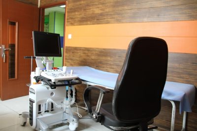
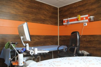
Eco cardiography
What is Eco cardiography?
Echocardiography, also known as an echocardiogram or cardiac echo test, is a procedure in which moving images of the heart are formed using sound waves. Echocardiography does not require hospitalization. It is a non-invasive procedure and does not cause any pain, unlike surgical procedures. Echocardiography is a diagnostic test used to assist your physician in identifying heart problems or assessing how your heart functions.
Echocardiography may be necessary in the following conditions:
- When you have abnormal heart sounds.
- When you experience unexplained chest pain.
- When you have a congenital defect.
- When you have had a heart attack.
- When you have rheumatic fever.
- When you experience shortness of breath during exercise, climbing stairs, or walking.

How is echocardiography performed?
The patient lies on their side or back on the bed. The doctor applies special gel to the probe and moves it over the chest area of the patient. Ultrasound waves create images of your heart and its valves. X-rays are not used in echocardiography. The heart's movements are observed on a video screen. A video or image can be obtained. During the test, the video can be viewed. This test is painless and has no side effects. The test usually takes less than 15 to 20 minutes. The doctor discusses the results with the patient. The timing of echocardiography: as advised by the cardiologist for hospitalized patients. What echocardiography shows: the size and shape of the heart, overall heart function, heart valve problems, and the presence of blood clots in the heart.
Responsible unit: Cardiologists (Dr. Nasir Moshtaqi, Dr. Khadijah Miaadi)
These services are only available to hospitalized patients in the hospital.


